

White velvet or orange smear is hyphae, yeast, and bacteria stacked.
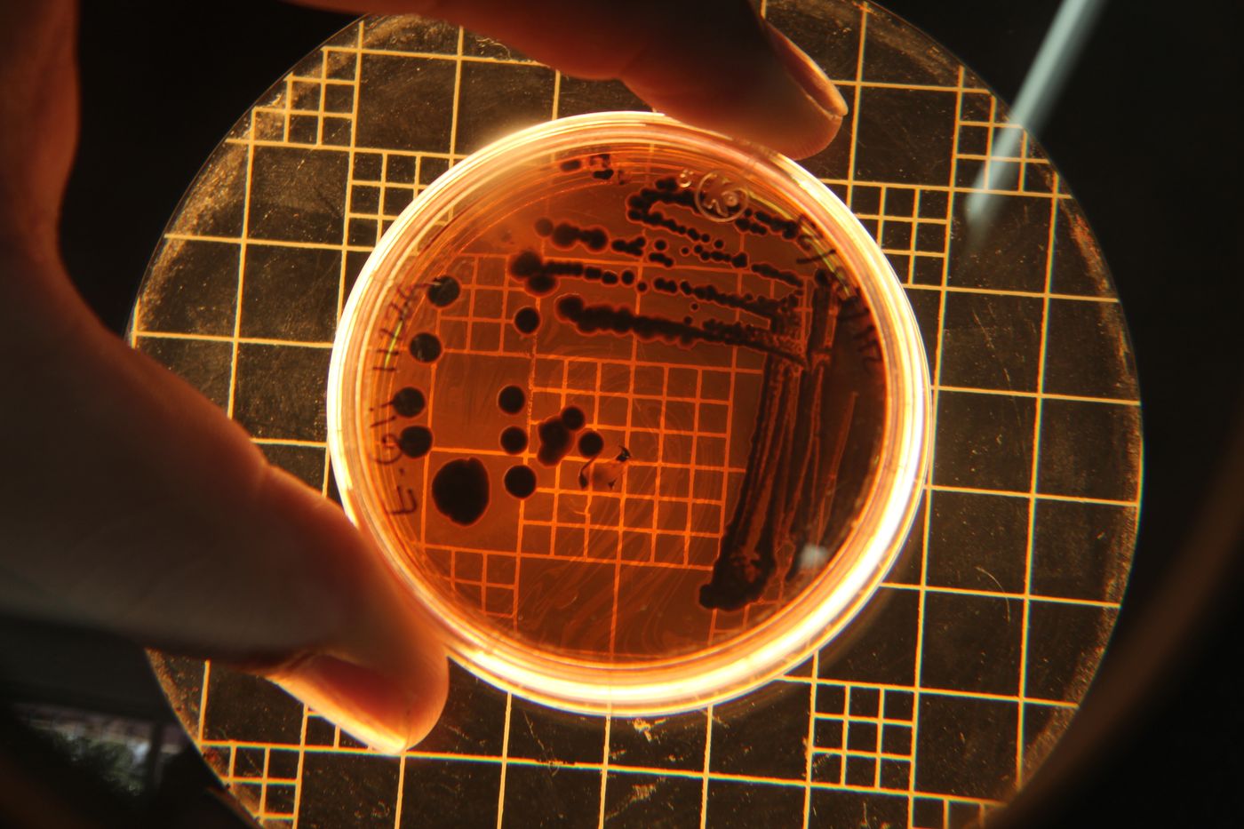
The point of a lens here is to see structure, not to baptise a new strain. A phone snapshot is not a species name.
What to look at, in order
- Naked eye. Frost, orange flecks, slime, a clean wrinkle, a vein that follows a pierce.
- Low power. Felted hyphae on a bloomy coat. A wet bacterial film on a smear. Dense threads along a blue vein.
- Higher. Oval yeasts. Bacteria as dust. You still will not get a Latin binomial from this kitchen.
A thin rind shaving in a drop of saline is enough to see whether you are looking at mycelium or a smear. That distinction already changes the note: bloomy versus washed, planned mold versus a wet failure.
A blue slice
Theatre, usefully: dense hyphae at the vein, softer paste where fat and protein have been taken apart. The marble is oxygen that was let in. Naming a new strain from that snapshot is another kind of failure.
Common mistakes
Calling every filament “Penicillium camemberti.” Posting a petri dish as if it were a finished identification. Scraping a soft cheese’s mold and declaring the interior safe because the surface “looked like Brie.” Using microscopy as a substitute for pH, weight, and a cold-chain log.
Sources
Legal ages, breeds, and cave rules are checked against official specifications or government pages. Sensory lines are editorial description, not lab results.
- Fox, P.F. et al., Fundamentals of Cheese Science (textbook, not a webpage) · Academic reference Mesophilic / thermophilic starter temperature bands; Curd cutting and whey expulsion
- FAO — Cheese making hygiene overview · FAO General cheesemaking process frame